|
RSD STRIATAL PHANTOM FOR SPECT/ PET/ MRI |
|
|
|
Fully
tissue-equivalent anthropomorphic phantom
This fully tissue-equivalent anthropomorphic
striatal phantom is designed for evaluation of quantitative striatal imaging
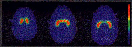
in humans using SPECT, PET or MRI. The phantom can be filled with radioactive
or MRI-signal-producing solutions. Ligands labeled with C-11, F-18 or I-123
are routinely used for evaluation of human neurodegenerative diseases such as
Parkinson's disease.
The phantom allows the effects of the imaging system on receptor
quantification to be investigated under conditions very similar to those in a
patient. It can be used to optimize the imaging system for patient imaging
and to examine many important issues related to receptor studies including:
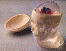
The phantom includes a transparent brain shell,
contained inside an accurately modeled human head, and a set of fillable
external markers. |
|
|
|
BRAIN SHELL The brain shell has five compartments which can be filled separately:
left and right nucleus caudate, left and right putamen, and the rest of the
brain. This allows different nucleus caudate to putamen ratios as well as
different striatal to background ratios to be obtained; this also permits differences
between left and right striatal activity to be examined. The volumes of the
nucleus caudate, putamen and the rest of the brain shell are 5.4, 6.0 and
about 1,250 ml, respectively. HEAD PHANTOM The modeled human head includes both the soft-tissue substitute and
the skull. The soft-tissue substitute is polyurethane, modified for tissue
equivalence, with a mass density of 1.10 g/cc. The narrow beam linear
attenuation coefficient measured at 140 keV (Tc-99m) is 0.160 cm-1 The
RSD bone substitutes closely meet the standards of the International
Commission on Radiation Units and Measurement (ICRU) Report No.44 (Tissue
Substitutes in Radiation Dosimetry and Measurement, 1989). The cortical bone
has a mass density of 1.86 g/cc. The narrow beam linear attenuation
coefficient measured at 140 keV is 0.280 cm-1. The nasal cavity
and maxillary sinuses are filled with foam with a mass density of 0.23 g/cc. FILLABLE EXTERNAL MARKERS A set of fillable capsules is provided to serve as external markers. Capsules
can be filled with a radioactive solution or CuSO4/NiCl2
and attached to the external surface of the phantom.
The phantom can then be imaged using any combination of SPECT, PET or MRI
modalities to compare image registration techniques. |
|
ORDERING INFORMATION |
|
|
MODEL |
DESCRIPTION |
|
RS-900T |
Striatal Phantom with Transparent Brain Shell and set of fillable
markers. |
|
RS-901T |
Transparent Brain Shell only with Striatum. |
© Elimpex-Medizintechnik, Spechtgasse 32, A-2340
Moedling, Austria
phone +43-2236-410450
fax +43-2236-410459