|
RSD ALDERSON HEART / THORAX PHANTOM FOR CARDIAC
SPECT/PET/MRI & MAMMOSCINTIGRAHY |
|
|
|
FEATURES:
|
|
This fully tissue-equivalent anthropomorphic heart/thorax phantom is
designed for cardiac SPECT/PET/MRI and mammoscintigraphy. The phantom can be
filled with radioactive or MRI-signal-producing solutions. Radiopharmaceuticals,
such as Tl-201, Tc-99m sestamibi, N-13 ammonia and F-18 FDG
(fluorodeoxyglucose), are routinely used to assess myocardium at risk,
infarct size and effectiveness of treatment. |
|
|
|
The phantom includes: 1.
Basic thorax
|
|
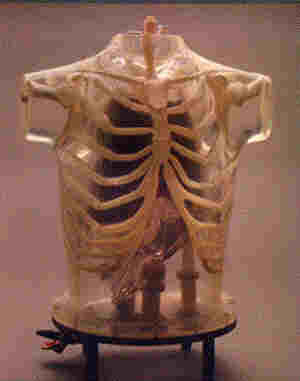
BASIC THORAX The
thorax is molded of polyurethane, modified for tissue-equivalence, with a
mass density of 1.10 g/cc. The narrow beam linear attenuation coefficient
measured at 140 keV (Tc-99m) is 0.160 cm-1. The
skeleton, embedded in the soft tissue, extends from the suprasternal notch
down to L2. The RSD materials closely meet the standards of the International
Commission on Radiation Units and Measurement (ICRU) Report No.44 (Tissue
Substitutes in Radiation Dosimetry and Measurement, 1989) for both the
cortical and spongiosa components of the human skeleton. The mass densities
of the cortical bone are 1.86 g/cc and of the spongiosa 1.16 g/cc,
respectively. The narrow beam linear attenuation coefficient for the cortical
component, measured at 140 keV, is 0.280 cm-1. The
volume of the thoracic cavity, when all organs (heart, lungs and liver) are
inserted, is about 8,200 ml. It is filled from the top, with either an inert
or a radioactive solution, to simulate the thoracic background. A valve
is installed at the base for conveniently draining the phantom. The residue
on the walls of the cavity and organs may be flushed by running water,
introduced by a hose attached to a fitting at the top of the phantom. A
smaller fitting at the top is an air-bleed, opened during filling and closed
during imaging. |
|
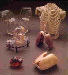
Disassembled Heart with Standard Defects |
|
HEART
An
accurately anatomic heart model is based on vacuum-formed shells. It was
designed using high resolution, contrast-enhanced, ultrafast CT data from a
normal patient, slightly modified to facilitate its use. The
left and right chambers are connected at the atrium region to make a single
compartment which can be filled and flushed independently using two ports
labeled HC (heart chambers). The right ventricle is slightly modified to
allow air to escape during filling. The myocardial wall (MW) has two ports,
flushing and independent filling. The volume of the heart chambers is 284 ml,
while the volume of the myocardial wall is 238 ml, without inserted defects. The
standard model includes three defects with volumes of 8.9, 13.5 and 41.7 ml,
respectively. Each of the defects can be filled separately. Defects
of different dimensions can be ordered at no added cost. A disassembled heart
is sent on request, so that dimensions of a special set can be established. Note
that different defects cannot be retrofitted in the assembled heart. |
|
|
|
Chest Overlay with
Breasts showing Tumors, Rods & Bending Fixture |
|
|

|
LUNGS There
are two models of lungs molded in hollow, vacuum-formed shells: 1.
Light-purple, non-perfusable lungs are molded in a syntactic foam with a mass
density of 0.30 g/cc. The narrow beam linear attenuation coefficient measured
at 140 keV is 0.043 cm-1 2.
Yellow, perfusable lungs are molded in an open-cell foam with a mass density
of 0.12 g/cc. The final mass density of 0.30 g/cc can be attained by varying
the volume of radioactive solution through a filling port at the top of each
lung shell. These
two lung pairs are provided, but extra sets of lungs can also be furnished
for work continuity. The volumes of the left and right lung shells are 907 ml
and 1,134 ml, respectively. LIVER
A liver
with a volume of 980 ml is included to evaluate the effect of its uptake on
quantitative myocardial imaging. It is a vacuum-formed shell, mounted on
perforated nylon tubes. The liver is filled with a radioactive solution and
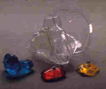
is about 5 mm from the heart. FILLABLE EXTERNAL MARKERS A set
of fillable capsules is provided to serve as external markers. Capsules can
be filled with a radioactive solution or with CuSO4/NiCl2 and
attached to the external surface of the phantom. It can then be imaged, using
any combination of SPECT/PET/MRI modalities to compare image-registration
techniques. THORAX OVERLAY, REMOVABLE BREASTS AND BREAST TUMORS The
thoracic phantom without overlay simulates an average male patient. The
overlay, with or without breasts, corresponds to a large female and a larger
male patient, respectively. Using these features it is possible to evaluate
the effect of additional attenuation and scatter on quantitative myocardial
imaging. The
volume of each vacuum-formed breast is 972 ml. A tumor filled with
radioactivity can be inserted to evaluate planar and tomographic imaging
techniques used for mammoscintigraphy. A set of breast tumors of 3, 5, 7, 9,
13 and 15 mm diameters is included. Tumors are supported by thin, threaded
nylon rods which pass through ports and are sealed by 0-rings. A bending
fixture is provided to permit the heated rod to be shaped to reach any part
of a breast. |
|
ORDERING INFORMATION |
|
|
MODEL |
DESCRIPTION |
|
RS-800T |
Heart/Thorax Phantom for Nuclear Medicine (Includes: RS-801 through
RS-810) |
|
RS-801 |
Thoracic Cavity with bottom plate |
|
RS-802 |
Non-Perfusable Lungs |
|
RS-803 |
Perfusable Lungs |
|
RS-804 |
Heart * |
|
RS-805 |
Liver Shell Only |
|
RS-806 |
Chest Overlay |
|
RS-807 |
Removable Breast with a set of tumors |
|
RS-808 |
Bending Fixture |
|
RS-809 |
Set of 25 threaded nylon tumor support rods |
|
RS-810 |
Set of 25 external fillable markers |
|
|
* with three hollow defects in myocardial wall. Standard sizes or to
customer specifications |
© Elimpex-Medizintechnik, Spechtgasse 32, A-2340
Moedling, Austria
phone +43-2236-410450
fax +43-2236-410459